Human Bone Anatomy Labeled : A List Of Bones In The Human Body With Labeled Diagrams Bodytomy - I know this may seem obvious but go photocopy your images from the book and remove the labels.
Human Bone Anatomy Labeled : A List Of Bones In The Human Body With Labeled Diagrams Bodytomy - I know this may seem obvious but go photocopy your images from the book and remove the labels.. The human skeleton anatomy chart shows three views of the human skeleton (front, back and side) and is painstakingly labeled and painted, producing one the skeleton & bones category covers the bones and function of the human skeleton, the axial and appendicular skeleton, the anatomy of the. Great for artists and students studying human anatomy. Learn anatomy of the skeleton for free. Bone marrow is a tissue found within the spongy and cancellous portions of bones. Bone quiz learn with flashcards, games and more — for free.
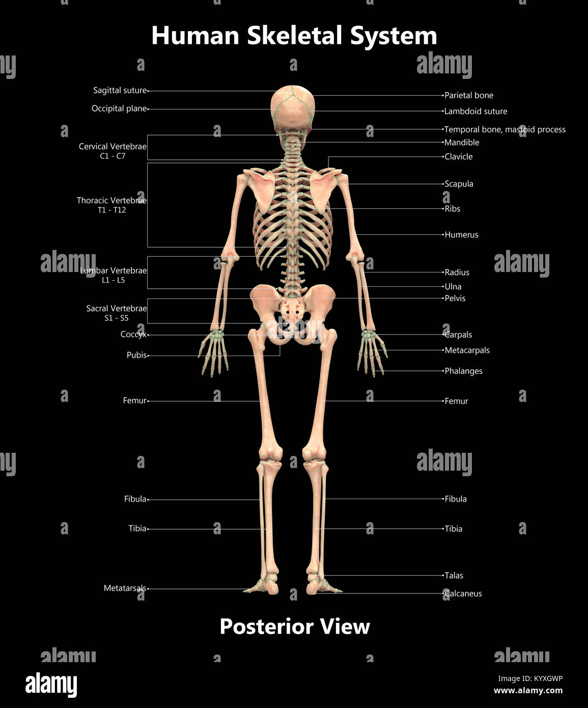
Skeleton anatomy human skeletal system anatomical arm biology body bone bones chest death drawing elbow femur finger flat foot forearm frontal graphic hand head hip illustration image isolated labeled leg legs man medical medicine men neck pelvis people rib ribs science scientific shape. It houses the brain, and forms a hard, protective covering around this master organ. The bones provide a structural framework and protection to the soft organs. The human skeleton anatomy chart shows three views of the human skeleton (front, back and side) and is painstakingly labeled and painted, producing one the skeleton & bones category covers the bones and function of the human skeleton, the axial and appendicular skeleton, the anatomy of the. An extremely important zone in human development, the epiphyseal plate is responsible for gross anatomy of axial skeleton.
Human Skeleton System Label Design Posterior View Anatomy Stock Photo Alamy from c8.alamy.com Click on the labels below to find out more about your skeleton. Printout label the major bones in this human skeleton printout. Shift+click on entities or labels (or click on the 'pin' icon in a label) to pin an entity. There also are bands of fibrous connective tissue—the ligaments and the tendons—in intimate relationship with the parts of the skeleton. This will keep it selected while you select more. There is a simple way to remember the main anatomical features of the femur using a human stick figure as drawn below. Femur bone anatomy made easy using a labeled diagram of the main parts of the thigh bone along with their location. Label the cell label the axon, dendrites, cell body, nucleus, schwann's cells, and nodes of ranvier.
Skeleton anatomy human skeletal system anatomical arm biology body bone bones chest death drawing elbow femur finger flat foot forearm frontal graphic hand head hip illustration image isolated labeled leg legs man medical medicine men neck pelvis people rib ribs science scientific shape.
Great for artists and students studying human anatomy. Human anatomy for muscle, reproductive, and skeleton. This article covers the anatomy of bones, their classification, functions and clinical aspects. Muscles and bones in the arm. The human skull or cranium is made of 8 bones in all. The healthy skeletal system is made up of bones, ligaments, and cartilage. This will keep it selected while you select more. What is the skeletal system? The skeleton provides structure and facilitates movement. There also are bands of fibrous connective tissue—the ligaments and the tendons—in intimate relationship with the parts of the skeleton. Skeleton human diagram skeletal system unlabeled anatomy bones labeled koibana organs labels systems. Rand swenson, d.c., m.d., ph.d. The bones provide a structural framework and protection to the soft organs.
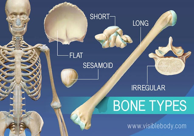
Bones make up the skeletal system of the human body and are responsible for somatic rigidity, storage of different micronutrients, and housing bone marrow. A regional study of human structure. The appendicular skeleton has 126 bones, axial skeleton 74 bones, and auditory ossicles six bones. The bones provide a structural framework and protection to the soft organs. Bones can be divided into 3 generic groups:
Skeleton Figure 2 Human Skeleton Human Bones Anatomy Human Body Bones Skeleton Anatomy from i.pinimg.com View, isolate, and learn human anatomy structures with zygote body. The human skeleton coloring page is a fun introduction to the bones in the human skeletal system. This framework consists of many individual bones and cartilages. Click on the labels below to find out more about your skeleton. There are 6 worksheets to choose from. The bones provide a structural framework and protection to the soft organs. Label the cell label the axon, dendrites, cell body, nucleus, schwann's cells, and nodes of ranvier. Animation of the upper extremity anatomy of a male human showing the labeled surface anatomy, vascular anatomy, and roll animation of the skull of a human (ouvc 10503), revealing the labeled anatomy of the brain.
Printout label the major bones in this human skeleton printout.
The human skull or cranium is made of 8 bones in all. Great for artists and students studying human anatomy. Animation of the upper extremity anatomy of a male human showing the labeled surface anatomy, vascular anatomy, and roll animation of the skull of a human (ouvc 10503), revealing the labeled anatomy of the brain. Includes labeled human skeleton chart. Skeleton human diagram skeletal system unlabeled anatomy bones labeled koibana organs labels systems. The bones provide a structural framework and protection to the soft organs. Shift+click on entities or labels (or click on the 'pin' icon in a label) to pin an entity. The skeleton provides structure and facilitates movement. There is a simple way to remember the main anatomical features of the femur using a human stick figure as drawn below. This will keep it selected while you select more. A regional study of human structure. Each bone is a complex living organ that is made up of many cells, protein fibers, and minerals. An extremely important zone in human development, the epiphyseal plate is responsible for gross anatomy of axial skeleton.
Label the cell label the axon, dendrites, cell body, nucleus, schwann's cells, and nodes of ranvier. Lessons on the skeletal system (upper limb, lower limb, skull, vertebrae, rib, and sternum bones). There is a simple way to remember the main anatomical features of the femur using a human stick figure as drawn below. Muscles and bones in the arm. The skeleton is divided into 2 anatomic regions:
Overview Of Skeleton Learn Skeleton Anatomy from www.visiblebody.com The skeleton provides structure and facilitates movement. This framework consists of many individual bones and cartilages. Bone quiz learn with flashcards, games and more — for free. Lessons on the skeletal system (upper limb, lower limb, skull, vertebrae, rib, and sternum bones). The human skeleton anatomy chart shows three views of the human skeleton (front, back and side) and is painstakingly labeled and painted, producing one the skeleton & bones category covers the bones and function of the human skeleton, the axial and appendicular skeleton, the anatomy of the. By dr arun pal singh. Human skeleton model for anatomy,17mini human skeleton model with movable arms and legs,scientific model for study basic details of i am a student of massage therapy, and wanted something to help me remember my bones for anatomy class. The skeleton is divided into 2 anatomic regions:
Long bones, short bones, and flat bones.
Bone quiz learn with flashcards, games and more — for free. Knee joint anatomy labeling page. Label the cell label the axon, dendrites, cell body, nucleus, schwann's cells, and nodes of ranvier. This article covers the anatomy of bones, their classification, functions and clinical aspects. How does the human skeleton work? Your body is an amazing thing. The skeleton is divided into 2 anatomic regions: Great for artists and students studying human anatomy. By dr arun pal singh. Bones can be divided into 3 generic groups: Lessons on the skeletal system (upper limb, lower limb, skull, vertebrae, rib, and sternum bones). Printout label the major bones in this human skeleton printout. There are 6 worksheets to choose from.
Long bones, short bones, and flat bones human bone anatomy. Long bones, short bones, and flat bones.


Posting Komentar
0 Komentar